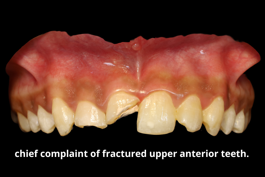
A 20-year-old male patient reported to Horizon Dental Clinic with the chief complaint of fractured upper anterior teeth. The patient was asymptomatic at the time of presentation and did not report any pain, swelling, or discomfort associated with the affected teeth.
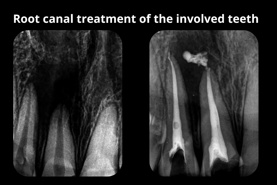
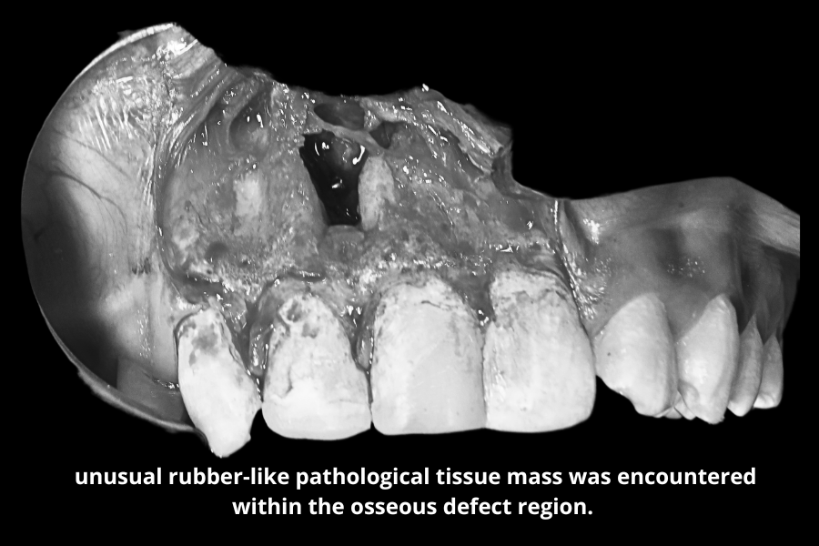
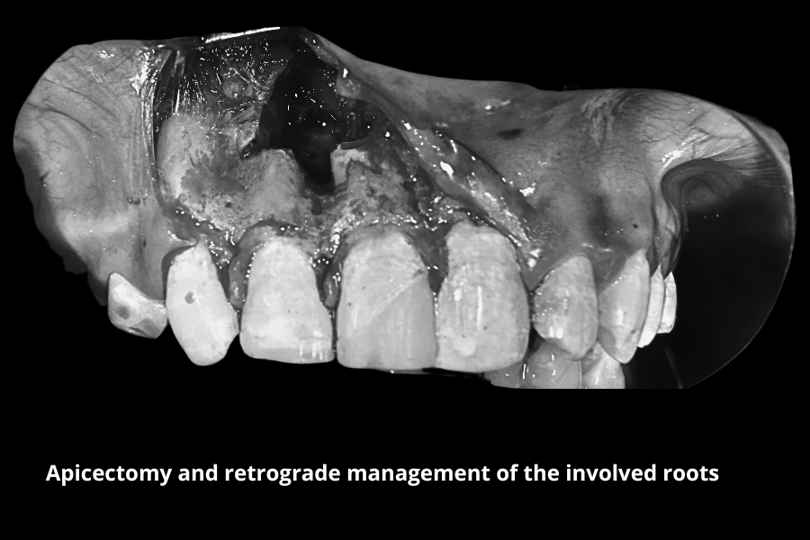
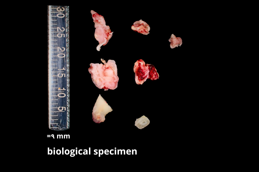
Clinical examination revealed fractured maxillary anterior teeth involving the central and lateral incisors. Radiographic assessment demonstrated a large periapical pathological lesion associated with the involved teeth, indicating chronic periapical pathology with significant bone involvement.
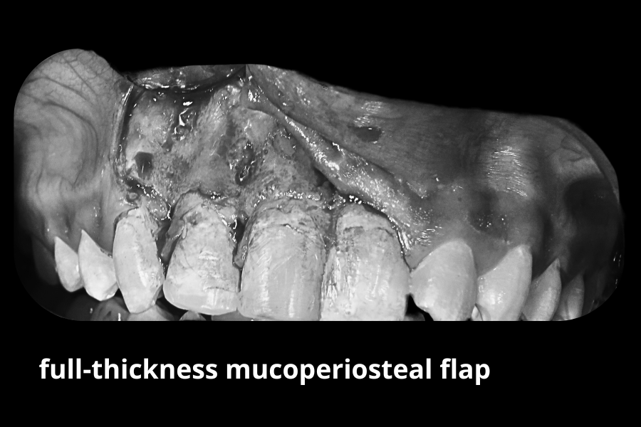
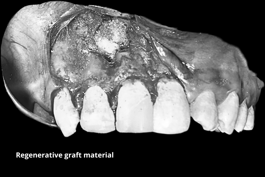
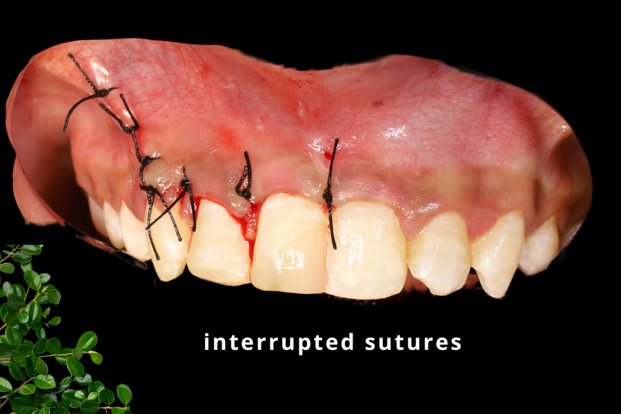
Considering the extent of the lesion and the strategic importance of the anterior teeth, a conservative tooth-preserving treatment approach was planned.
The proposed treatment protocol included:
The procedure was planned under local anesthesia with the objective of eliminating pathology, preserving natural dentition, and promoting optimal hard tissue healing.
Following administration of local anesthesia, a full-thickness mucoperiosteal flap was carefully reflected to access the periapical lesion. During surgical exploration, an unusual rubber-like pathological tissue mass was encountered within the osseous defect region.
The surgical site was extensively irrigated to eliminate residual debris, inflammatory by-products, and diluted pathological contaminants from the defect area. Apicectomy and retrograde management of the involved roots were completed as planned.
Regenerative graft material was placed within the osseous defect to support bone healing and tissue regeneration.
The excised biological specimen was preserved and submitted to a pathology laboratory for histopathological evaluation and further investigation to confirm the definitive nature of the lesion.
The surgical site was then repositioned and secured using interrupted sutures under adequate hemostasis.
The patient was kept under periodic post-operative observation and demonstrated satisfactory healing without pain, swelling, or secondary complications. Sutures were removed after 7 days, and the surgical site exhibited favorable soft tissue healing with stable post-operative recovery.
This case highlights the importance of comprehensive radiographic diagnosis and multidisciplinary surgical-endodontic management in the preservation of compromised anterior teeth associated with extensive periapical pathology. Careful surgical debridement combined with regenerative management can significantly improve healing outcomes and help maintain natural dentition in young patients.
Open for Appointments